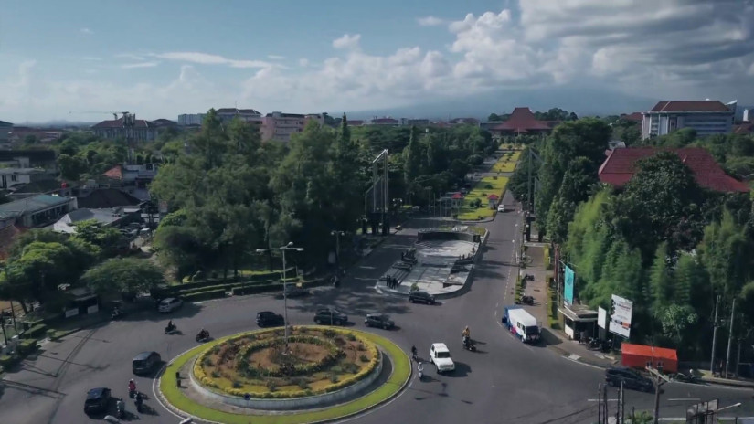
UGM memiliki 18 fakultas dan 2 sekolah yang menyelenggarakan program sarjana, sarjana terapan, pascasarjana, profesi, spesialis, serta program internasional. Ragam bidang ilmu di UGM dikelompokkan menjadi 4 klaster, yakni kesehatan, sains dan teknologi, agrokompleks, dan sosiohumaniora.

Merupakan wujud nyata kontribusi UGM terhadap Indonesia. Setiap tahunnya, ribuan mahasiswa diterjunkan ke seluruh penjuru tanah air untuk mengabdi dan berpartisipasi dalam pengembangan masyarakat.
Merupakan jaringan organisasi pendidikan formal, nonformal, dan informal yang ada, yang dimobilisasi untuk menyampaikan pendidikan untuk pembangunan berkelanjutan (ESD) kepada masyarakat lokal dan regional.

Perhelatan akbar tahunan International Conference and Annual Meeting Association to Advance Collegiate Schools of Business (ICAM AACSB) telah berlangsung pada 14-17 April 2024 di Atlanta, Georgia, Amerika Serikat. Perhelatan prestisius tersebut dihadiri ratusan pimpinan Fakultas dan Sekolah dari seluruh dunia, baik anggota AACSB yang belum terakreditasi maupun yang telah terakreditasi. Sebagai sekolah bisnis yang telah […]
Sampah plastik masih menjadi persoalan dalam pengelolaan sampah di Indonesia yang sampai saat ini sulit terselesaikan. Pasalnya komponen plastik menjadi jenis sampah yang sulit dihancurkan. Menurut Kementerian Lingkungan Hidup dan Kehutanan, terdapat total timbunan sampah di Indonesia yang mencapai 17,4 juta ton hingga tahun 2023, adapun sampah plastik yang menyumbang sekitar 17,29% dari total sampah […]
Kagama.co pernah menulis kiprah Yahyawan Triyana, alumnus Teknik Pertanian, Fakultas Teknologi Pertanian UGM. Dalam tulisan tersebut Yahyawan mengaku cukup sederhana beralasan ingin kuliah di Fakultas Teknologi Pertanian UGM: ingin belajar teknologi tepat guna di jurusan yang dinaungi Fakultas Teknologi Pertanian (FTP) UGM. Cukup sederhana. Tapi siapa tahu, kesederhanaan itulah yang pada akhirnya membuatnya memiliki bermacam […]
Pelopor perguruan tinggi berkelas dunia yang unggul dan inovatif, mengabdi kepada kepentingan bangsa dan kemanusiaan.

Mencetak mahasiswa berwawasan global dan berintegritas yang dilandasi nilai-nilai Pancasila.
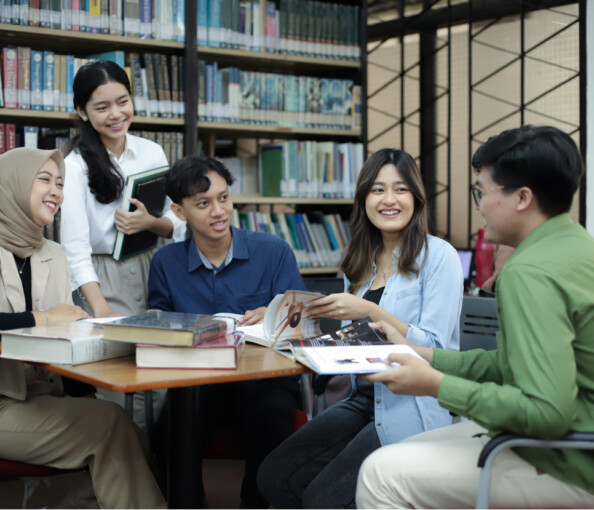
Alumni UGM tersebar di berbagai wilayah, berkiprah di tingkat nasional dan global.