
UGM memiliki 18 fakultas dan 2 sekolah yang menyelenggarakan program sarjana, sarjana terapan, pascasarjana, profesi, spesialis, serta program internasional. Ragam bidang ilmu di UGM dikelompokkan menjadi 4 klaster, yakni kesehatan, sains dan teknologi, agrokompleks, dan sosiohumaniora.


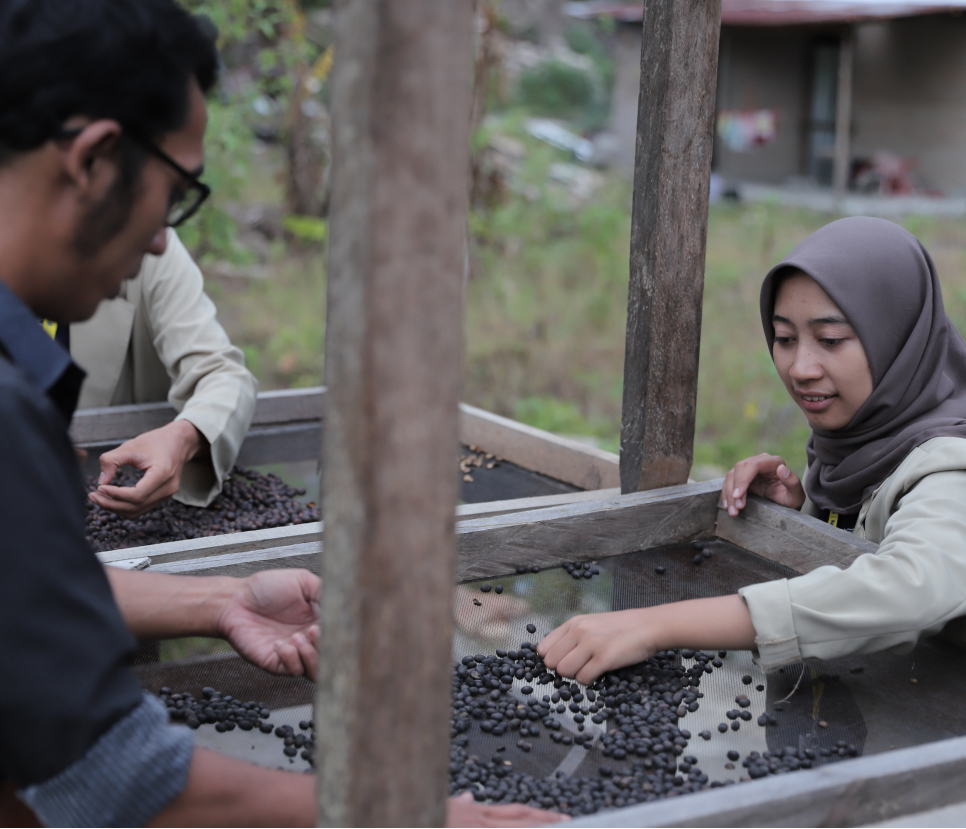
Merupakan wujud nyata kontribusi UGM terhadap Indonesia. Setiap tahunnya, ribuan mahasiswa diterjunkan ke seluruh penjuru tanah air untuk mengabdi dan berpartisipasi dalam pengembangan masyarakat.
Merupakan jaringan organisasi pendidikan formal, nonformal, dan informal yang ada, yang dimobilisasi untuk menyampaikan pendidikan untuk pembangunan berkelanjutan (ESD) kepada masyarakat lokal dan regional.
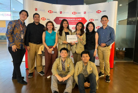
Tim Mahasiswa dari FEB UGM kembali menorehkan prestasi di tingkat nasional dengan menjadi juara pertama dalam HSBC Business Case Competition 2024 pada 20-24 April 2024 lalu di Jakarta. Tim beranggotakan Nathaniel Christanto Kuniardi dan Gildas Sebastian Satriatama Akuntansi dari prodi Akuntansi, Adena Laksita Paramesti dari prodi Manajemen, dan Cynthia Aretha Pratiwi A dari Ilmu Ekonomi […]
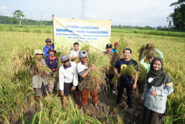
Varietas padi Gamagora hasil riset Pusat Inovasi Agroteknologi (PIAT) UGM berhasil dipanen petani yang tergabung dalam Gabungan Kelompok Tani (Gapoktan) Makmur Sejahtera, Sentolo, Kulon Progo. Panen padi Gamagora yang perdana ini dilakukan dalam demplot seluas 1 ha dan terbagi tiga titik. Panen varietas padi Gamagora pada Selasa (23/4) memperlihatkan hasil rata-rata 7,52 ton/ha untuk wilayah […]

Kamu berencana kuliah di Fakultas Ekonomika dan Bisnis? Jika iya, sebaiknya kamu harus tahu lebih jauh soal prodi yang ditawarkan di Fakultas Ekonomika dan Bisnis Universitas Gadjah Mada (FEB UGM). Sebagai informasi, FEB UGM merupakan sekolah bisnis pertama di Indonesia yang berhasil mendapat akreditasi internasional the Association to Advance Collegiate Schools of Business (AACSB), lembaga […]
Pelopor perguruan tinggi berkelas dunia yang unggul dan inovatif, mengabdi kepada kepentingan bangsa dan kemanusiaan.

Mencetak mahasiswa berwawasan global dan berintegritas yang dilandasi nilai-nilai Pancasila.
Alumni UGM tersebar di berbagai wilayah, berkiprah di tingkat nasional dan global.