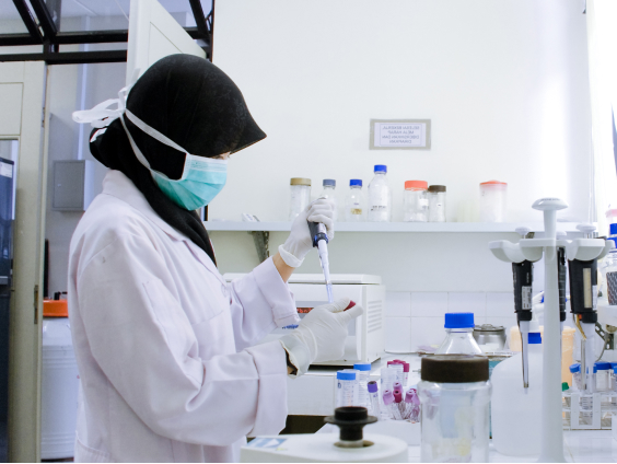

UGM memiliki 18 fakultas dan 2 sekolah yang menyelenggarakan program sarjana, sarjana terapan, pascasarjana, profesi, spesialis, serta program internasional. Ragam bidang ilmu di UGM dikelompokkan menjadi 4 klaster, yakni kesehatan, sains dan teknologi, agrokompleks, dan sosiohumaniora.


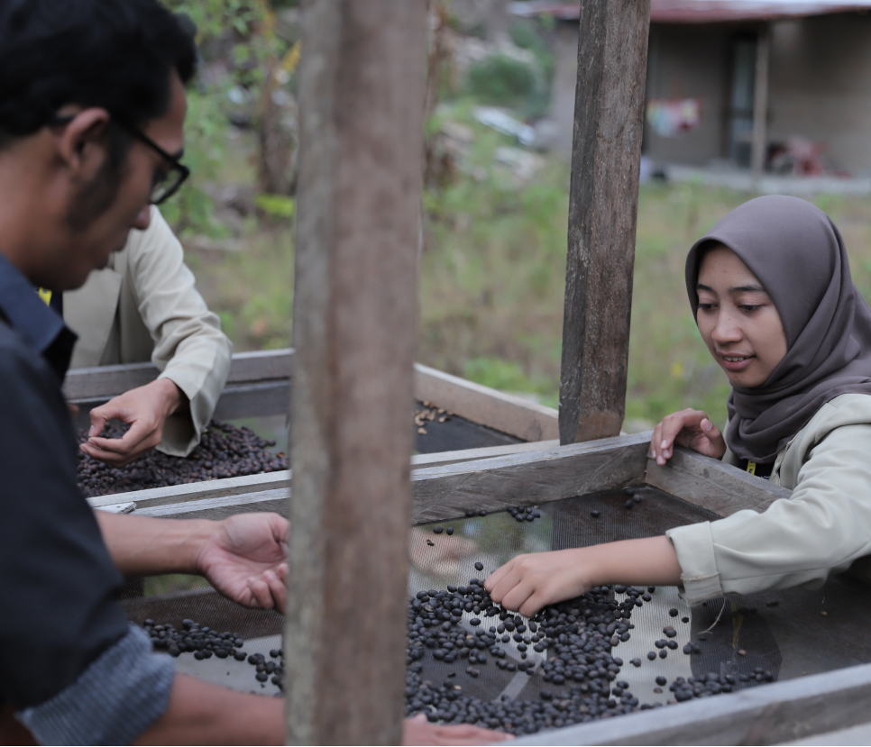
Merupakan wujud nyata kontribusi UGM terhadap Indonesia. Setiap tahunnya, ribuan mahasiswa diterjunkan ke seluruh penjuru tanah air untuk mengabdi dan berpartisipasi dalam pengembangan masyarakat.
Merupakan jaringan organisasi pendidikan formal, nonformal, dan informal yang ada, yang dimobilisasi untuk menyampaikan pendidikan untuk pembangunan berkelanjutan (ESD) kepada masyarakat lokal dan regional.

Universitas Gadjah Mada senantiasa berkomitmen untuk memberikan kualitas pendidikan terbaik, serta meningkatkan kredibilitas sebagai world class university. Tahun ini, sebanyak 25 bidang ilmu UGM tercatat dalam peringkat QS World University Rankings (WUR) by subject 2024, tiga diantaranya ada di Fakultas Ilmu Sosial dan Ilmu Politik (Fisipol) UGM. Adapun tiga program studi yang masuk dalam daftar ranking […]

Berpulangnya komedian Babe Cabita akibat penyakit anemia aplastik menyebabkan nama penyakit ini menjadi semakin dikenal luas oleh masyarakat. Seiring dengan informasi soal penyakit ini, muncul sebuah konten di platform salah satu media sosial yang menyebutkan salah satu merek obat sakit kepala yang disebut dapat menyebabkan anemia aplastik. Sontak, masyarakat menjadi heboh, karena obat sakit kepala […]

Ingin berkarier sebagai dokter gigi? jika iya, maka kamu harus tahu profesi dokter gigi dapat dicapai dengan kuliah di prodi Sarjana Kedokteran Gigi selama empat tahun dengan gelar Sarjana Kedokteran Gigi (S.K.G). Selanjutnya kamu harus menempuh pendidikan koasistensi selama 1,5 tahun untuk mendapat gelar profesi dokter gigi (drg). Namun menjadi dokter gigi tidak hanya sebatas […]
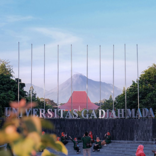
Pelopor perguruan tinggi berkelas dunia yang unggul dan inovatif, mengabdi kepada kepentingan bangsa dan kemanusiaan.

Mencetak mahasiswa berwawasan global dan berintegritas yang dilandasi nilai-nilai Pancasila.
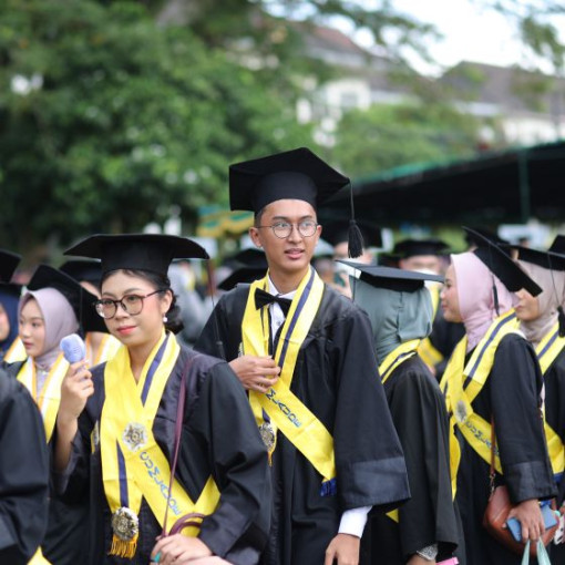
Alumni UGM tersebar di berbagai wilayah, berkiprah di tingkat nasional dan global.